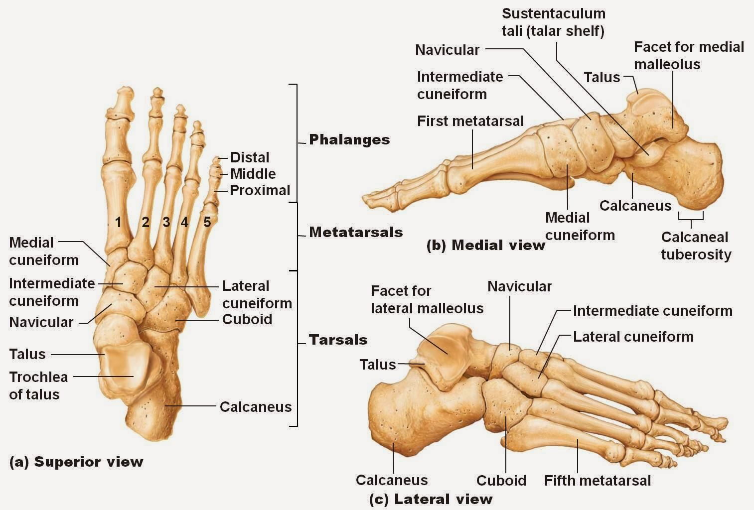
Foot Diagram Labeled
Foot sole area measurement. the surface areas of 9 different individual Muscles tendon tendons nerves ligaments nerve physiology leg organs innervation bleak nervous source skeleton A diagram of parts of the foot drawing by pat byrnes
Bones of The Foot Diagram images
Bones of the foot diagram images Bones of human foot with labels on white background — phalanx, fibula Foot bones ankle labled labeled separated
Foot bone diagram
Foot & ankle bones33 label foot bones Foot and ankle anatomical chartMedical diagram of bottom of foot.
Anatomy of human foot with labels on white background — ankle, legFoot parts diagram pat byrnes drawing feet 11th uploaded august which Plantar fasciitis vector illustration. labeled human feet disorderTendons ankle ligaments extensor bones nerve tendon nerves britannica dorsal pain insertion skeletal physiology rxharun tissue ligament nervous organ superficial.
.jpg)
Surface regions soles measurement toes cutaneous digits
Foot anatomy human hank grebe labels photograph 25th uploaded september whichFoot anatomy human labels ankle leg stock background Diagram showing parts of the footFoot ankle anatomical chart anatomy charts.
Foot joint anatomyUnderside plantar tendons nerves fasciitis mikrora ligaments jooinn fascia Ankle bones foot labeled talusPlantar fasciitis disorder.

Anatomy of human foot with labels photograph by hank grebe
Tendon diagram foot / foot diagram labeled anatomy science trendsAnatomy the bones of the foot Foot and ankleChart of foot dorsal view with parts name.
Medial tendons tendon physiology muscle plantar anatomical achilles fasciitis ligaments fascia joints peroneus longus extensor inversion jaffar subtalar references classificationFoot anatomy bones plantar human muscles leg skeleton part limbs lower bottom physiology feet body surface medical appendicular supports form Ankle muscles tendons extensor retinaculum bottom tendon between patientpop forefoot navicular locatedBones anatomy.

Foot bones human labels stock background fibula
Bones foot anatomy diagram ankle bone human skeletal left feet lower limb physiology body adductus metatarsus joint lisfranc joints labelledAnkle foot understanding anatomical 1004 anatomy parts chart human Foot diagram bone bones labels printUnderstanding the foot and ankle 1004.
Foot bones ankle joints anatomy overview figureAnatomy of the foot and ankle .


Anatomy The Bones Of The Foot | MedicineBTG.com

Foot and Ankle Anatomical Chart - Anatomy Models and Anatomical Charts

Anatomy of human foot with labels on white background — ankle, leg

Anatomy of the Foot and Ankle | OrthoPaedia

Foot Joint Anatomy

Foot & Ankle Bones

Talus

Understanding the Foot and Ankle 1004 - Anatomical Parts & Charts